MCQ ON HUMAN EYE class 11 for NEET | HUMAN EYE class 11 | MCQ HUMAN EYE with Answer | Check the below NCERT MCQ question for class 11 Biology based on NCERT the with Answers.
MCQ ON HUMAN EYE class 11 for NEET
MCQ on HUMAN EYE class 11 Biology with answers were prepared based on the latest pattern.We have provided class 11 Biology MCQs question with Answers to help students understand the concept very well.
MCQ ON HUMAN EYE is useful for NEET / CSIR / UGC / CBSE / ICSE / AIIMS / EXAM / AFMC EXAM / STATE LEVEL MEDICAL EXAM 2022-23 , 2023-24
INTRODUCTION:
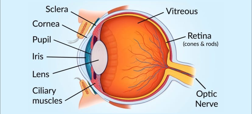
Our paired eyes are located in sockets of the skull called orbits. The adult human eye ball is nearly a spherical structure. The wall of the eye ball is composed of three layers.
The external layer is composed of a dense connective tissue and is called the sclera.
The anterior portion of this layer is called the cornea.
The middle layer choroid contains many blood vessels and looks bluish in colour.
Choroid layer is thin over the posterior to 2/3rd of the eyeball, but it becomes thick in the anterior portion to form the ciliary body.
The ciliary body itself continues forward to form a pigmented and opaque structure called the Iris which is the visible coloured portion of the eye.
The eye ball contains a transparent crystalline lens which is held in place by ligaments attached to the ciliary body.
In front of the lens the rounded by the Iris is called the pupil.
The diameter of the Pupil is regulated by the muscles fibres of iris.
MCQ ON HUMAN EYE 11 for NEET
1. The wall of human eye is nearly a spherical structure.The wall of the eye ball is composed of ……
layers
(a) two
(b) three
(c) four
(d) all the above
Ans (b) three
2. The external layer of eye is composed of a dense connective tissue and it is called
(a) sclera
(b) cornea
(c) choroid
(d) ciliary body
Ans. (a) Sclera
3. The anterior portion of the layer of human eyes is called
(a) sclera
(b) cornea
(c) ciliary body
(d) all the above
Ans. (b) cornea
4. The middle layer of human eye contains many blood vessels and looks bluish in colour
(a) cornea
(b) sclera
(c) choroid
(d) ciliary body
Ans.(c) choroid
5. The visible coloured portion of the eye is
(a) Iris
(b) sclera
(c) choroid
(d) cornea
Ans.(a) Iris
6. In front of the lens the aperture surrounded by the Iris is called
(a) pupil
(b) cornea
(c) iris
(d) all the above
Ans.(a) pupil
7. The diameter of the pupil is regulated by
(a) cornea
(b) sclera
(c) muscles fibres of iris
(d) retina
Ans.(c) muscles fibres of iris
8. The inner layer of eye is a retina which contains 3 layers of cells from inside to outside
(a) ganglion cells
(b) bipolar cells
(c) photoreceptor cells
(d) all the above
Ans.(d) all the above
9. There are two types of photoreceptor cells found in eye.
(a) rods
(b) cones
(c) both a and b
(d) none of the above
Ans. (c) both a and b
10. The daylight vision and colour vision are functions of
(a) rods
(b) cons
(c) both a and b
(d) none of the above
Ans. (b) cones
11. Twilight (scotopic) vision is the function of
(a) rods
(b) cons
(c) both a and b
(d) none of the above
Ans.(a) rods
12. The rods contain a purple red protein called rhodopsin or visual purple which contains derivative of
(a) Vitamin A
(b) Vitamin B – compex
(c) Vitamin C
(d) Vitamin D
Ans . (a) Vitamin A
13. Photo receptor cells are not present in that region and it is called
(a) blind spot
(b) optic nerve
(c) fovea
(d) macula lutea
Ans.(a) blind spot
14. It is a thind out portion of the retina where only the cons are densely packed.
(a) optic nerves
(b) blind spot
(c) fovea
(d) all the above
Ans. (c) fovea
15. The space between the cornea and the lens is called
(a) aqueous chamber
(b) vitreous chamber
(c) both a and b
(d) blind spot
Ans.(a) aqueous chamber
ALSO READ:-
● YOU CAN WATCH BIOLOGY SIR Youtube channel
16. The space between the lens and the retina is called
(a) aqueous chamber
(b) vitreous chamber
(c) both a and b
(d) blind spot
Ans.(b) vitreous chamber



Leave a Comment