
Heart: anatomy, physiology, location, disease and treatment
Vertebrate have single heart. it is hollow muscular organ composed of cardiac muscles fibre which are striated muscles fibre in nature. It acts as pumping organ of blood vascular systems. it receives blood from and pump blood to the various organs and tissues of the body. for this purpose it undergoes spontaneous, rhythmical and ceaseless contraction (beat) throughout the Animals life.
The hearts is divided internally into intercommunicating chambers the number which varies is in different vertebrate groups. the heart in all vertebrate has 1 or 2 auricles (atria) and 1 or 2 ventricles. these auricles and ventricles chambers are known as true Chambers.
The heart of lower vertebrate as additional Chambers namely sinus venosus and conus arteriosus or bulbous arteriosus or truncus arteriosus. these Chambers are termed as accessory Chambers.
Heart: anatomy, physiology, location, disease and treatment
During the course of development in higher vertebrate the persistent portions auricles and ventricles are retained. however this get complicated by incorporating several valves inside them and becoming compartmentalized. In such cases the accessory chambers sinus venosus and conus arteriosus are displaced from their original position and are actually incorporated into the right wall of Atrium and ventral aorta respectively.
◆ Follow me on YouTube
◆ VISIT ON OUR YOUTUBE CHANNEL BIOLOGY SIR FOR MORE VIDEO
The study of hearts is known as cardiology. Composed of two word kardia and logos. Kardia is word meaning hearts and logos means study so cardiology is study of heart.
Table of Contents
Fishes heart and single type of circulation
● Fishes :- A fish heart handles only deoxygenated blood (venous) and is often called Venus heart. It is two chambered having single auricle and single ventricle and the accesory Chamber sinus venosus and conus arteriosus are present.
Fishes heart and single type of circulation
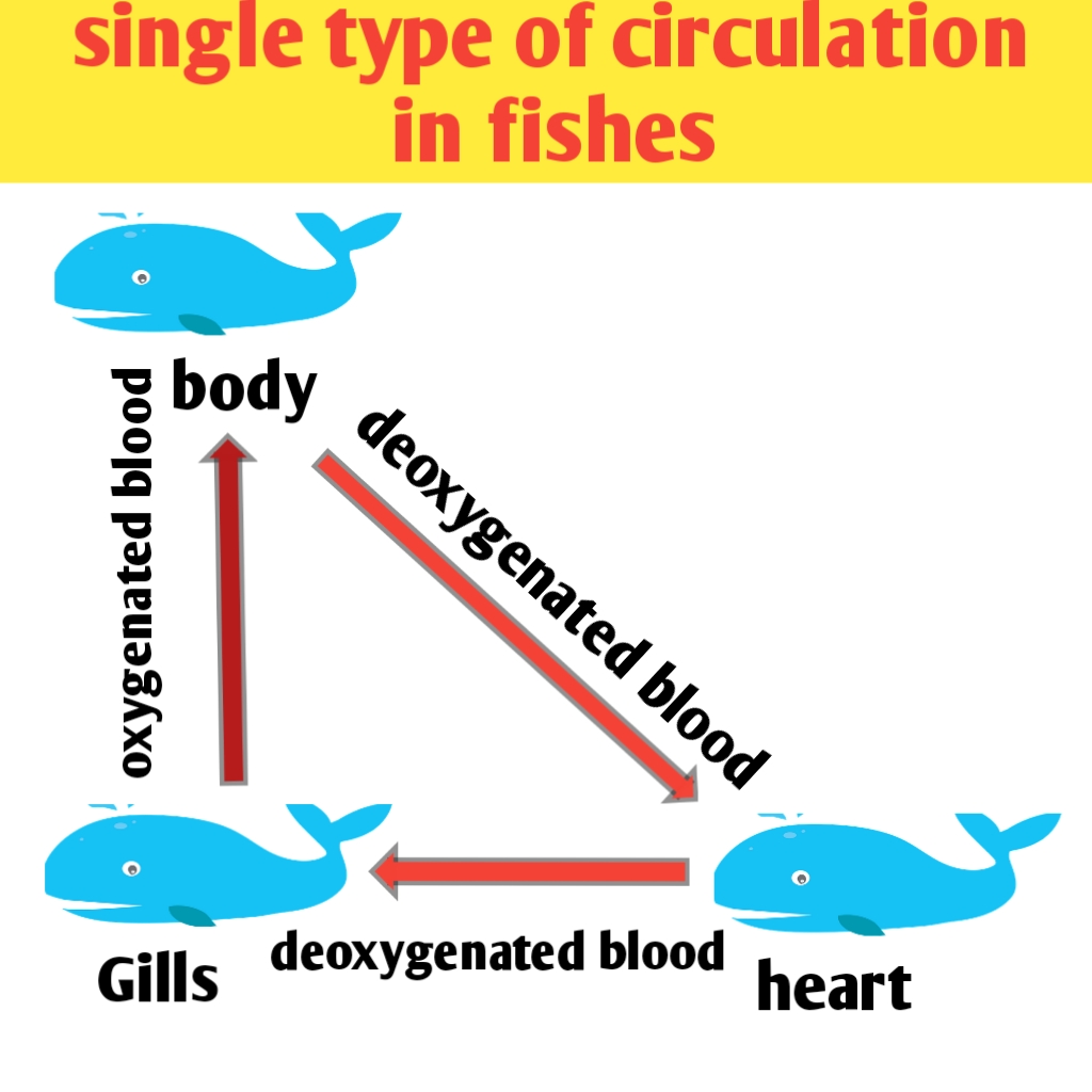
The hearts receives deoxygenated blood from all over the body except the gills into the Sinus venosus and Pump it via auricle, ventricle and conus arteriosus into a short artery, the ventral aorta,or which distribute into the gills by breaking of into capillaries, here,the blood picks up oxygen.
The oxygenated blood from the gills flows into the dorsal aorta whose branches distribute it to the rest of body by splitting into capillaries. Here, the blood distribute Oxygen and become deoxygenated. The deoxygenated blood returns to the heart through the veins. Since the blood passes through the hearts only once in a complete circuit around the body, the fishes are said to have a single circulation.
◆ Follow me on YouTube
◆ VISIT ON OUR YOUTUBE CHANNEL BIOLOGY SIR FOR MORE VIDEO
Advantage of single circulation in fish
An advantage of single circulation is that the entire body receives oxygenated blood. A disadvantage is that narrow Gill capillaries slow down the blood flow and the body receives blood at a low pressure. this slow the rate of oxygen supply to the cells and limit the metabolic rate that fish can attain.
Amphibian heart and double type of circulation
Amphibian heart receives both deoxygenated (Venus) and oxygenated (arterial) blood, that’s why it it is often termed as arteriovenous heart. It is three chambered having two (right and left) auricles and a single undivided ventricles, two acessories Chambers sinus venosus and truncus arteriosus are also present.
Amphibian heart and double type of circulation
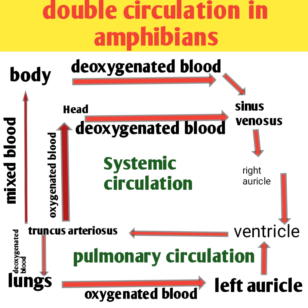
The right auricle receives deoxygenated blood from the body via sinus venosus and the left auricle receive oxygenated blood from the lungs. Both the samples of blood passes into the ventricles, where mixing of two occurs. The truncus arteriosus supplies mix blood to the various part of the body.
The amphibian have Double circulation as the blood passes through the heart twice in each complete circuit around the body. The course of blood from the heart to the lungs and back to the hearts is known as pulmonary circulation.
The flow of blood from the hearts to the body and head and back to the heart is termed systemic circulation.
Advantage of double circulation in Amphibia
An advantage of double circulation is that the blood supply to the body is also pumped by the hearts. This raises the blood pressure and accelerates the supply of Oxygen and food to the body cells.
Use of undivided ventricle in Amphibia
What is the use of having and inefficient and undivided ventricle in which oxygenated and deoxygenated blood get mixed up ? Most Amphibians spend a good deal of time underwater where air breathing is not possible. Their undivided ventricles can route the blood from the body past the lungs and can send it to the skin for further gaseous exchange with the water. In amphibians much gas exchange occurs in the skin an added advantage for submerged animal.
Reptiles heart and blood circulation
Reptilian heart also receives both oxygenated and deoxygenated blood and is, thus arteriovenous heart. It is incompletely four chambered having two auricles and a single partialy divided ventricles only one accessory chamber name sinus venosus is present.
Circulation of blood in amphibia
The right auricle receives deoxygenated blood from the body via sinus venosus, and the left auricle receives oxygenated blood from the lungs, both the samples of blood pass into the ventricles, which sends deoxygenated blood to the lungs via pulmonary arch and oxygenated blood to the rest of the body via pair of systemic arch. Little mixing of blood occurs in the ventricle because of valve in it.
Thus the ventricle is functionally divided, though not structurally, reptiles also have a partial double circulation.
Birds and mammals hearts
Avian and mammalian heart is four chambered, having right and left auricles and right and left ventricles. the right auricle receive the deoxygenated blood from the body and send it to the right ventricle that Pump It to the lungs via pulmonary arch for oxygenation.
Double circulation in fishes and mammalia
The left auricle receives oxygenated blood from the lungs and send it into the left ventricle which Pump It to the body through a single aortic arch. Thus the oxygenated and deoxygenated blood remain fully separate, And there is a complete double circulation.
Advantage in mammalian heart
This is the main advantage in the Avian and mammalian heart over the amphibian and reptile hearts and there are no accessory Chambers. Completely four chambered heart of birds and mammals supplies more oxygen to tissues because there is no mixing of oxygen rich blood and oxygen poor blood.
◆ Follow me on YouTube
◆ VISIT ON OUR YOUTUBE CHANNEL BIOLOGY SIR FOR MORE VIDEO
This is necessary because these vertebrates need more oxygen per gram of body weight then other vertebrate of equal size. they are endotherms and use heat released from metabolism to warm the body.
Advantage of single Aortic Arch in mammalia
Blood leaves the ventricle for supply to the body by two aortic arches in reptiles and by a single aortic arch in birds and mammals. Blood passing through two Arctic arch es loses pressure faster than when it flows through one aortic arch.
Higher blood pressure in birds and mammals means faster circulation and quicker supply of Oxygen and food to the tissues and Rapid removal of wastes material from them. Complete separation of pulmonary and systemic circulation allows the two circulation to half different blood pressure according to the need of the organ they supply.
Mammalian (Human) heart
appearance and position
The heart is a hollow, muscular organ, roughly of size of one’s fist (12 × 9 cm). Its average weight is about 300 gram in male and about 250 gram in females. It attain the adult weight at the age of 17 to 20 years. The heart is a reddish brown in colour and somewhat conical in form.
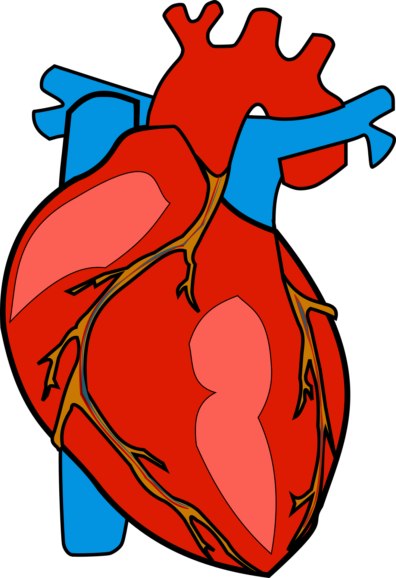
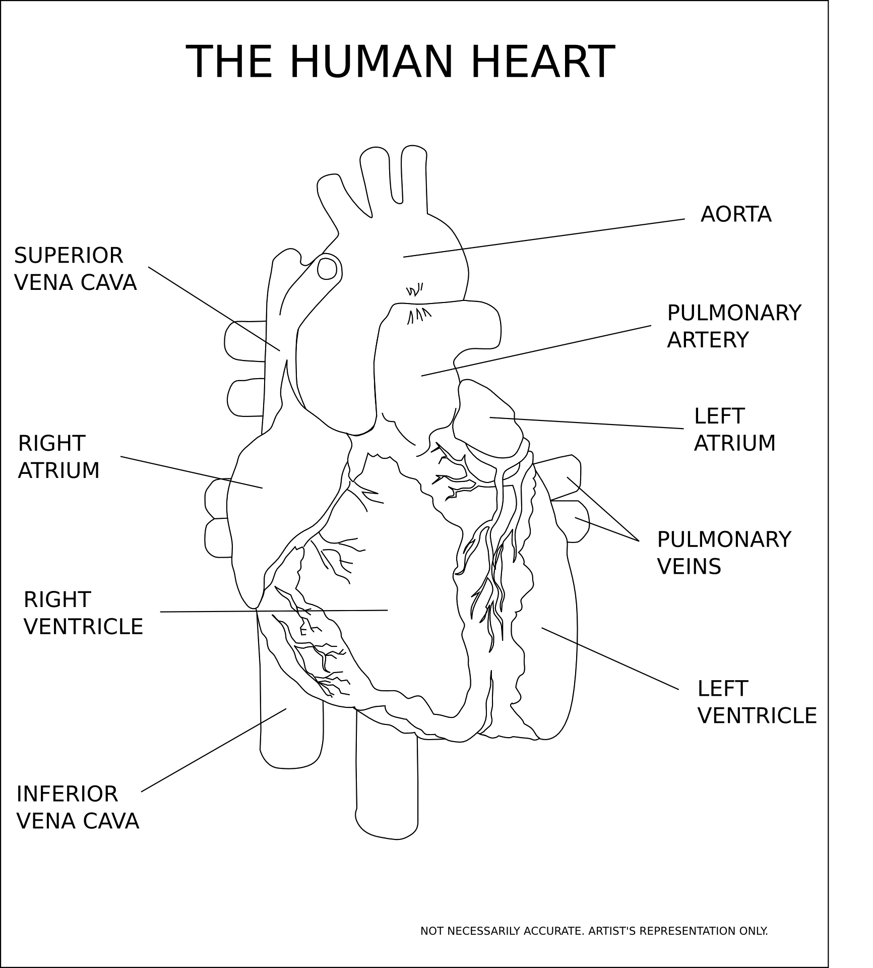
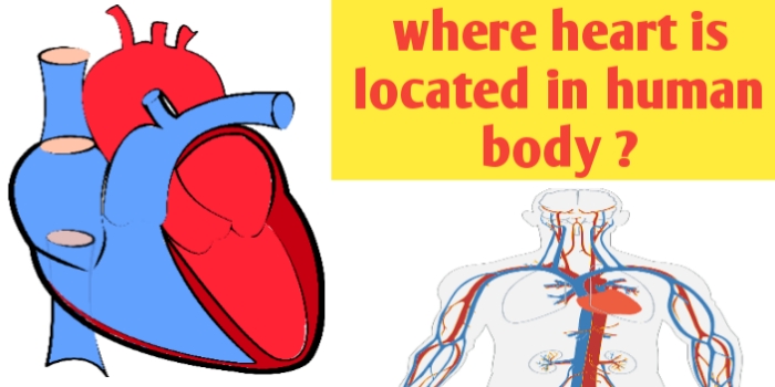
Human heart image or picture
Where is is heart located in human body ?
It is located almost in the middle of the thoracic cavity close to its front wall and between the lungs. Its broad base faces upward and backward. Its narrow apex is directed downward, forward and slightly to the left, and raised on the diaphragm, is heart located left or right ? It is slightly located left from the midline of the body and two third of mass is in left side, average 15 centimetre left to midline of the body.
Human heart structure and anatomy
Is heart a muscle or organ ?
Is heart a muscle or organ ? Human hearts is actually a muscular organ. An organ is a group of tissues that work together to perform a specific biological function. In the case of humans heart, this function is pumping blood throughout your body. Additionally, the heart is largely made up of a type of muscle tissue called cardiac muscle.
Protective covering of heart
The hearts is enclosed in a tough, two layered sac, the pericardium, comprising inner visceral pericardium attached to the heart and Outer parietal pericardium. The two layers are continuous at the roots of great vessels, and have between them a potential space known as pericardial cavity. This cavity contains of upto 50 ml of pericardial fluid.
The latter is secreted by the pericardium itself, and occurs as thin film between the two pericardial layers. This fluid keep the heart moist, allows free movement and reduce the friction between the heart wall and the surrounding tissues when the heart beats. The pericardium protect the heart from mechanical injury and checks it over stretching or overfilling with blood.
Heart Anatomy (internal and external structure)
Heart anatomy is comprise of of external and internal structure
External structure of heart
The heart is a double Pump, It is divided by the septa into two halves, the right and left. each half consists of two communicating Chambers upper a smaller auricle or atrium and lower larger ventricle. Thus the heart has four Chambers.
Human heart four chambered and anatomy
● Auricles :- the auricles form a small upper part of the heart they are demarcated externally from the ventricles by an irregular groove called the coronary sulcus. Each auricle is produce into a flap, the auricular appendix, that project over the corresponding ventricle.
● Ventricles :- the ventricles form a large lower part of the heart. the two ventricles are the demarcated externaly from each Other by an oblique groove termed as interventricular sulcus. This groove contains coronary blood vessels the right ventricles does not reach the apex.
heart four chambered and internal structure
Human heart is consist of four chamber namely right auricle, left auricle, right ventricle and left ventricle.
● Auricles :- the auricles act as collecting chamber for the blood returning to the heart, they have thin walls because they have to force the blood into the ventricles that lies just below them. the inner surface of auricle is a smooth but for a network of low ridge, the musculipectinati, in the region of auricular appendages.
The two auricles are separated from each other by a partition known as interauricular or interatrial septum. The septum has an oval thin area called fossa ovalis. It marks the position of an opening, the foramen ovale, between the two auricles in the foetus.
● Ventricles :- the ventricles act as distributing Chambers for the blood reaching from the Atria. They have thicker walls than the auricles have, and the wall of left ventricle is about three time as thick as that of the right ventricle. This is because the left ventricle has to pump the blood to the farthest end of the body, where is the right ventricle has to send the blood to the lungs which lie nearby.
Correspondingly the blood entering the Aortic Arch from the left ventricle is at much higher blood pressure than the blood entering the pulmonary arch from the right ventricle.
The inner surface of the ventricles is raised into a network of low muscular ridge known as columnae corneae or trabeculae carnea. And a few large conical muscular elevation term as musculi papillares or papillary muscles. The to ventricles are separated from each other by a thick curved partition known as interventricular septum.
Great blood vessels, Aperture and Valves
The blood vessels that enter or leave the heart are known as great blood vessels
Vena Cava great blood vessel
the right auricles receives two large veins : superior vena cava and inferior Vena cava. The superior Vena cava brings deoxygenated blood from the head and upper region of the body. The inferior Vena cava returns the deoxygenated blood from the lower region of the body.
The opening of the inferior Vena cava is Bordered by membrane, falciform fold, which is a remnant of the foetal Valve of Eustachius. The right auricle also receive a small coronary sinus. It returns deoxygenated blood from the heart wall. Its opening is guarded by a small fold known as valve of Thebesius.
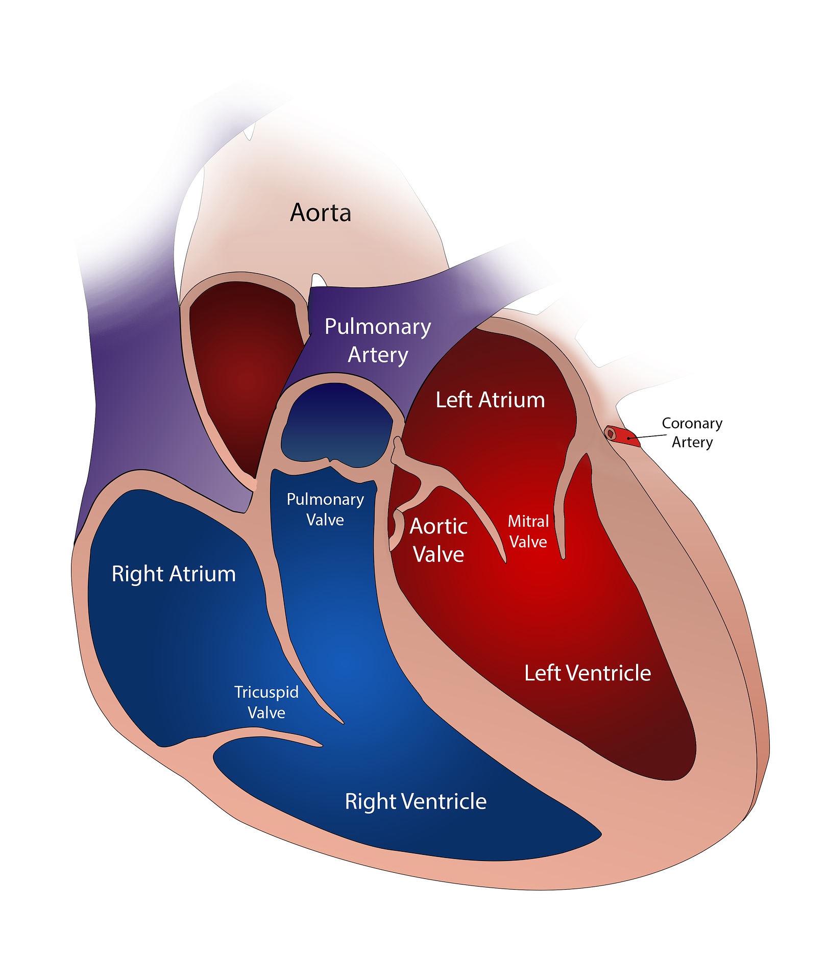
Right A-V Aperture and Tricuspid Valve
the right auricle opens into the right ventricle through a wild passes the right auriculoventricular or A-V aperture. This aperture is guarded by one way valve called the tricuspid valve.
The tricuspid valve consists of three membranous flaps which are attached to the margin of the right auriculoventricular aperture but project freely into the ventricle below. Their free lower edges are fixed to the papillary muscles of the right ventricle by a number of tough,white cords known as the chordae tendineae. The chordae tendineae check the pushing of the flaps into the auricles during the contraction of the ventricle.
Pulmonary Arch and Semilunar valves
from its upper left corner the right ventricles give off a large blood cells called the pulmonary arch or pulmonary aorta. It divides into right and left pulmonary arteries that carry deoxygenated blood to the lungs for oxygenation.
At the base of Pulmonary arch 3 membranous pocket shaped flaps, the semilunar or S – L valves, set in a ring with their cavities directed away from the ventricles. They prevent the return of the blood to the ventricles.
pulmonary Veins
the left auricle receives four pulmonary veins, two from each side. They bring oxygenated blood from the lungs and they have no valves
Left A – V Aperture and Bicuspid Valve
the left auricle opens below into the left ventricle by a large passage known as the left auriculoventricular or A – V aperture. This aperture is guarded by one way valve known as bicuspid or mitral valve. It consists of two flaps only. The upper edges of these flaps are attached around the left auriculoventricular aperture, while their lower edges project freely into the left ventricle. The free edges of these flaps are connected by chordae tendineae to the papillary muscles of the left ventricle.
Systemic aorta and Semilunar valves
at its upper right angle, the left ventricle give off a large blood vessels called the systemic aorta. The latter has three regions, ascending aorta, arch of aorta and descending aorta. At the base of ascending aorta are three membranous pocket shaped semilunar valve with their cavities directed away from the ventricle. These valve check the return of the blood to the ventricle.
Just beyond the semilunar valve in the ascending aorta give off right and left coronary arteries that supply blood to the hearts wall.
The arch of aorta gives of three large arteries: brachiocephalic, left common carotid and left subclavian arteries. The descending arch extends through the trunk and supplies oxygenated blood to the various part axcept the lungs.
Sphincters of great veins
at their entry into the atria, the venae cava and Pulmonary vein have rings of muscles which contract and close of these vessels during atrial contraction. This prevent Reflux of the blood into them.
Ligamentum Arteriosum
a ligament known as ligamentum arteriosum connect the pulmonary aorta with the systemic aorta. it is the remnant of an embryonic channel between the two aortae.
Histology of human heart
The heart wall consist of three layers, inner endocardium, middle myocardium and external epicardium.
◆ endocardium: it comprises and endothelium of squamous cells resting on a thin layer of loose connective tissues
◆ myocardium: it consists of cardiac muscle cells are found in layers that run in complex spiral manner. The cells are of two types, first one is contractile cells and second one is impulse generating and conducting cells. There is a central fibrous skeleton of dense connective tissues. The Skeleton serves as support and as site of origin and insertion of cardiac myocytes.
◆ Epicardium: it is the visceral pericardium. it consists of an outer simple squamous epithelium also known as mesothallium resting on a thin layer of connective tissues.
Human heart physiology and working
The heart pump the blood to all the parts of body. it pumps out over 7000 litres of blood daily. force that propel the blood comes from the contraction of cardiac muscles, which form the hearts wall. the cardiac muscle cells are striated, packed with contractile protein myofilament and branched forming a network. As a result the cardiac muscles cells contract into two dimension, wringing the blood out of the heart with great force.
The heart contract more than 2.5 billion times in a life time. to pump out blood the heart Chambers undergo alternate contraction known as systole and relaxation known as diastole. The contraction of a chamber decreases its volume and forces the blood out of it, where as it relaxation brings it back to its original size to receive more blood.
Cardiac cycle or heart cycle
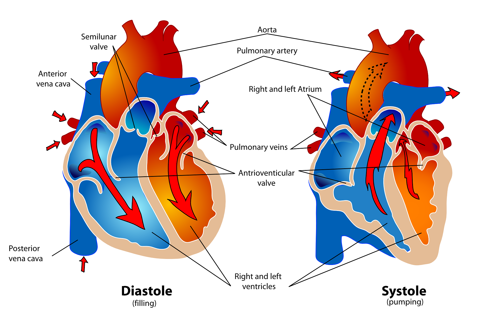
the completion of one heart beat is known as heart or cardiac cycle. Cardiac cycle consists of a regular sequence of three events: (1) auricular systole, (2) ventricular systole and (3) joint diastole or complete cardiac diastole (relaxation of both auricles and ventricles). These events are repeated in a cyclic manner during each Heart beat and make the blood flow through the heart Chambers in a specific manner and direction.
Cardiac cycle systole and diastole
Joint diastole
Joint Diastole during this phase blood from the Great veins and coronary sinus flows into the auricles and some blood also passes from the auricles into the respective ventricles. this flow of blood is called not by contraction of any part of the heart but simply by the fact that pressure in the heart is less than that in the great veins and the A-V valves are open.
Auricular Systole
◆ Auricular Systole: contraction of the auricles drives most of their blood into the respective ventricles which are still relaxed (ventricular diastole). During auricular systole all the blood does not return to the Great veins as the contraction of the auricles begins at their upper ends and passes toward the ventricles, forcing the blood into the latter.
More over during auricular contraction the sphincters at the opening of great vein close, roots of great veins are compressed, blocking their opening. Also the end of auricle systole, there start relaxation of auricles known as auricular diastole and contraction of the ventricles known as ventricular systole simultaneously.
Ventricular Systole
◆ Ventricular Systole: As the contraction of the ventricles begins, the pressure of blood in them almost immediately Rises above that in the auricles. With this pressure the flaps of the A-V valves are pushed up and meet to close the A-V apertures. This tight closure of the A-V valves at the start of the ventricular contraction produce the first heart sound known as “lub“.
The semilunar valves of great arteries remain closed for a moment, so that the blood is blocked on all sides and its pressure Rises with further contraction of the ventricles. When the pressure in the ventricles exceeds that in great arteries, the semilunar valve are pressed, the apertures they guard open, and the blood spurts into great arteries. This Is the End of the ventricular systole.
Now the ventricles start relaxing (ventricular diastole), the auricles are still undergoing diastole, at this time all the Chambers of heart are in diastole known as joint diastole.
As the ventricular diastole progress, the pressure in the ventricles Falls below that in the great arteries, the flow of blood from the ventricles stops and the semilunar valves close to check the backflow of blood from the great arteries to the ventricles. The semilunar valves close as they get filled with blood trying to return and meet together, closing the outlets of great arteries.
The closure of the semilunar valves at the start of the ventricular diastole for the second hearts sound known as “dup”. The sound “lup” is lower pitch and last longer than the sound “dup“. The ventricles remained as closed chamber for a moment because pressure in them is still higher than that in the auricles and this keep the A-V valves closed.
Duration of heartbeat
one complete heart beat or cardiac cycle last for 0.8 seconds. Its three events namely, auricular systole, ventricular systole and joint diastole take 0.1 seconds, 0.3 seconds and 0.4 seconds respectively.
Nourishment of heart (coronary circulation)
A pair of coronary artery arises from the systemic aorta the just beyond its semilunar valves and break up into capillaries to the vascularise the heart wall. Blood is returned from the heart wall by number of coronary veins that join to form of coronary sinus.
The latter opens into right auricles. the arteries and veins of heart wall form the coronary circulation. The blood filling the heart Chambers is quite far from most of tissues forming the thick heart wall. More over food and oxygen requirements of the heart wall are high in view of its constant beating. Any damage to the coronary vessels will disrupt the supply of blood and food and oxygen to the heart tissues. It may, therefore proof very serious.
Efficiency of heart
The hearts keeps on beating throughout life without fatigue because it rest and works for equal durations. The contraction of the heart Chambers is followed immediately by relaxation but relaxation is not followed At Once by the next contraction. For the next contraction starts there is a resting or recovery period in which no activity is seen. The recovery period is 0.4 second is as long as the contracting time (0.1 + 0.3) second in which the hearts shows activity and does work.
Humane heart beat
◆ heart beat definition: the are spontaneous and rhythmic contraction and relaxation of the heart to pump out and receive blood to and from the body is known as heart beat.
Heart beat types
◆ Heart beat types: the Heartbeat is of two types neurogenic and myogenic
Neurogenic heart beat
◆ Neurogenic heart beat: it is initiated by a nerve impulse emanating from a nerve situated near the heart. it is shown by the hearts of most orthopods and some annelids.
myogenic heart beat
◆ myogenic heart beat: it is initiated by a patch of modified heart muscles itself without requiring and external stimulation. It is exhibited by the hearts of mollusca and vertebrates.
A heart removed from frogs body continuous to beat for a long time if emerged in well oxygenated ringer solution that supplies essential element and nutrients. This shows that the Heart beat has its mechanism in the heart wall itself. And is independent of nervous and endocrine system. these systems however to control the rate of heartbeat.
Mammalian heart removed from the body does not continue to beat for so long because it is more sensitive to temperature and an adequate oxygen supply.
mechanism of heart beat
◆ mechanism of heart beat: the Heartbeat results from a wave of electrical depolarization (action potential) called as cardiac impulse, that originates in and is conducted along the tracts of specialised cardiac muscles fibre term Purkinje fibers. These tracts are collectively referred to as the nodal tissue or Purkinje system.
morphology of nodal tissues
◆ morphology of nodal tissues: the nodal tissue consists of Sinuauricular node commonly known as S-A node, auriculoventricular node (A – V) node, bundle of His or auriculoventricular bundle (A -V) bundle, right and left bundle branches and purkinje fibres.
The S – A node is located in the Wall of the right auricle slightly below the opening of the superior Vena cava. This is the place where the Sinus venosus is incorporated in the embryo.
The A-V node lies in the wall between the right auricle and right ventricle. The bundle of His originates from A-V node the passes through the interauricular and interventricular septa, and divides into the right and left bundle branches, one going into the wall of each ventricle. In The Wall of the ventricles the two branches break up into a large number of Purkinje fibre that are distributed to the entire musculature of the ventricles.
working of nodal tissue
◆ working of nodal tissue: the S-A node has unique property of self excitation, which enables it to act as “pacemaker” of the heart. It is continuously initiates a wave of contraction which spreads over both the auricles more or less simultaneously along the muscles fibre that and fan out from the pacemaker.
The auricular contraction cannot pass over the ventricles because the musculature of the auricles and ventricles are not continuous and are separated by an atriventricular septum of fibrous connective tissue, the annular pad, in mammals.
The auricular contraction how ever stimulates the A-V node which generates a wave of contraction that passes over both the ventricles simultaneously along the bundles of His and its ramifications. The ventricular contraction begins at the apex of the heart and passes quickly towards the origin of the pulmonary and systemic arches.
At the A-V node the impulse are delayed for about 0.1 second to ensure that the auricle will contract first and empty fully before the ventricles contract.
The S-A node sets up cardiac impulses 72 times per minute. It means heart beat 72 times in one minute. The S-A node not only acts as pacemaker but also stabilizes the basic rhytm at which the heart beats.
The impulses which travel through cardiac muscles during the cardiac cycle produce electrical currents the latter are conducted through body fluids to the body surface, where the current can be deleted by electrodes placed on the skin and recorded as an electrocardiogram (ECG).
Control of heart beat
◆ Control of heart beat: the rate of the Heartbeat is however controlled by the nervous and endocrine systems.
nervous control of heart beat
◆ (1) nervous control of heart beat: Sinuauricular node received from the brain two sets of nerve fibre, sympathetic that stimulates the S-A node and accelerate the heartbeat and parasympathetic or vagal that inhibit the S-A node and retard the heartbeat. Sensory fibre extend from the stretch receptors present in the Wall of the aortic arch, carotid sinuses and vena cava to the cardiovascular centre in the medula oblongata.
The impulses received from the Aortic arch and carotid sinuses decrease the heart rate where as the impulse is from the vena cava increases the heartrate.
The integrated activity of inhibiting and accelerating effects occurring in the medulla oblongata control the heart rate the heart does always work against a break at any given time the heart rate is compromise reached by opposing action of two sets of nerves.
hormonal control of heart beat
◆ hormonal control of heart beat: hormones from the medulla of adrenal glands epinephrine and norepinephrine accelerate the Heartbeat at the time of emergency.
Body temperature also affects the pacemaker. just 1 Degree Celsius rise in temperature increases the heart rate by about 10 beats per minute. This is why our pulse rate is much higher in fever. The heart rate also increases with exercise, this is an adaptation that enables the circulatory system to provide additional Oxygen and food needed by muscles during hard work
Human heart rate
● Heart rate: human heart beats about 72 times per minute in adult person at rest. This is called the heart rate. It varies in different species. Generally the smaller animals have higher higher rates than the longer ones. The reason for this is that a smaller animals is usually have higher metabolic rate and their heart must be beat faster to supply the required amount of nutrients and oxygen to the tissues. The heart rate increases during exercise, fever and emotion such as anger and fear.
Heart rate in infant or newborn baby
Your baby’s heart rate should be between 90 to 110 beats per minute at 6 to 7 weeks. By the ninth week your baby’s Heartbeat should reach up to 140 to 170 beats per minute.
maximum heart rate
You can calculate your maximum heart rate by subtracting your age from 220, for example if you are 45 years of old subtract 45 from 220 to get a maximum heart rate of 175, so maximum heart rate during exercise is 175 beats per minute.
Normal heart rate
Normal heart rate
Normal heart rate for adult ranges from 60 to 100 beats per minute. Generally lower heart rate at rest implies more efficient fitness for example a well trained athlete might have normal resting heart rate closer to 40 beats per minute.
Heart rate in human in different age group
Infants belonging to 6 month have heart rate 120 -160 beats per minute, Toddler of 2 years have heartrate 90 – 140 bpm, pre-schooler children have heart rate 80 -110 beats per minute, children belongs to school age have heart rate about 75 -100 beats per minute, adolescent age have heart rate about 60 to 90 beats per minute and in adult have heart rate about 60 to 100 beats per minute
heart rate in animals
Heartrate in cattle is 60-90 bpm, in calves is 100 -120 bpm, in horse 28 – 42 bpm, in foal 70 -80 bpm, in sheep 68-90 bpm, in pig 60 – 90 bpm, in dog 70 -130 bpm, in cat 110 -130 bpm ,in elephant 30 bpm and in chicken 200 – 400 bpm.
Human heart pulse rate
The flow of blood from the heart into the artery create pulse. The ventricle pump out blood when it contracts and turn out no blood when it relax. Each ventricular system forces blood into the arteries which already contain some blood. The arteries expand to receive the incoming blood during diastole the elastic recoil of the expanded part of artery process the blood into the next part of the artery which in turn distends.
A wave of destination passes along the arteries following is ventricular systole this wave of destination is known as arterial pulse. It can be felt in superficial arteries such as those in the wrist, neck, temples and ankles. It is generally felt by placing fingers over the radial artery near the wrist. Since each Heartbeat generates one pulse in the arteries the pulse rate per minute indicate the heart rate. So pulse rate and heart rate are identical which is being 72 per minute
So normal range of pulse rate is about 72 per minute, and it will be increased during exercise when we feel emotions such as angry and fear, and Pulse rate is more in infants or newborn baby.
Human heart sounds
There are two types of sound produced during the cardiac cycle “lup” and “dup”
first heart sound lub or “lup”
● first heart sound “lup”: it may be likely to represent by the word lub or “lup”, are spoken very softly. It is low pitch not very loud and of long duration. It is produced by the simultaneous tight closure of the tricuspid and bicuspid valves at the start of ventricular contraction. And it last for 0.15 seconds and its principal frequencies are in the range of 25 to 45 cycles per second
second heart sound “dup”
● second heart sound “dup”: it has been known as “dup”. It is higher pitched, louder and of short duration. It is produced by the closure of the semilunar valves at the start of ventricular relaxation. It lasts for 0.1 seconds and its principal frequency is 50 cycles per second.
Both the heart sounds can be heard by placing ear or better stethoscope reciever on the left side of the chest wall at the fourth intercostal space.
heart sound Murmur
◆ heart sound Murmur: Certain disease such as syphilis and rheumatic fever, many damage heart valves. Defective or damaged heart valves produce during their closure an abnormal sound reffered to as murmur. It is detectable as a hissing sound when a stream of blood squirts backward through a valve. Some people are born with heart murmurs.
Cardiac output ( heart output)
The volume of blood pumped by the left ventricle into the systemic circuit per minute is called heart output or cardiac output. It depends on two factors: heart rate (pulse) and stroke volume, the amount of blood pumped by the left ventricle each time it contracts.
Thus 72×70 or 5040 ml roughly 5 litres is the heart or cardiac output. This is only a little less than the total amount of blood about 6.8 litres present in the body. During activity, the heart output increases to supply more food and oxygen to muscles. Mild exercise such as walking, raises the heart output to about 11 litres. During vigorous exercise, it may rise to 25 litres, and to 40 litres in a trained athlete.
What is blood pressure ?
Blood pressure is the pressure against the wall of the blood vessels produced by the discharge of blood into them by contraction of the left ventricle.
What is blood pressure ?
Role of blood pressure
The blood pressure is high in the arteries gradually drops in the arteriols and capillaries, and become very low in the vein. It is this marked gradient of blood pressure that keep the blood flowing from the arteries through the capillary to the vein. Fluids always flow from area of higher pressure to area of lower pressure.
Systolic and diastolic pressure
All main arteries have almost the same average pressure throughout their length. The pressure in the arteries Rises and fall alternately due to heartbeat. When the heart contracts the blood is forced into the arteries which expand to accommodate the blood.
The temporary rise in blood pressure during the contraction of the heart is known as systole pressure and the temporary fall in blood pressure during relaxation of the heart is known as diastolic pressure.
Determination of blood pressure
Blood pressure is generally measured by determining the millimetres of Mercury Hg displaced in pressure gauge called sphygmomanometer. It Express as ratio of systolic pressure over the diastolic pressure. For healthy resting adult person the normal range of blood pressure is 120/80 mm Hg. Where systolic pressure is 120 mm Hg and diastolic pressure is 80 mm Hg.
Pressure drops with increasing distance from the heart the brachial artery in the arm a little above the elbow is used for measuring blood pressure. Systolic pressure shows the force with which the left ventricle pushes blood into the Aortic arch. The diastolic pressure indicates the elasticity of the blood vessels and is useful in diagnosing hardening of arteries or is strain on their walls.
The difference between systolic pressure and diastolic pressure is known as the pulse pressure which is about 40 mm Hg for normal person. pulse pressure may be also defined as variation occurring in the artery during the cardiac cycle.
Hypertension
Hypertension is persistent rise in blood pressure called high blood pressure or Hypertension. It results from narrowing of arterial lumen and reduced elasticity of arterial wall in old age. It can cause rupturing of capillaries and it is also known as silent killer.
Factors affecting blood pressure
The blood pressure is influenced by many factor such as force and rate of Heartbeat, resistance offered by arteries and capillaries to the flow of blood, viscosity of blood and volume of blood pumped out at each Heartbeat and increase in any of these factors can raise the blood pressure. Age is also affect blood pressure.
Hypotension
Fall in the arterial blood pressure is known as low blood pressure or hypotension. It may result from chronic expansion of loss of blood in human or failure of pumping action of heart it may cause fainting.
Human heart diseses
Heart disease is also known as cardiovascular disease many disease affect the blood vessels and the heart these include hypertensive heart diseases, coronary heart disease, rheumatic heart, heart block and stroke.
Hypertensive heart disease includes arteriosclerosis, atheroma and hypertension
Arteriosclerosis heart disease
◆ Arteriosclerosis: arteriosclerosis is the hardening of arteries and arterioles due to thickening of fibrous tissues and the consequent loss of elasticity. It causes hypertension or high blood pressure in this disease calcium salt precipitate with the cholesterol of the formed or forming plaque.
This calcification ultimately makes the wall of arteries stiff and rigid. Such arteries lost the property of distension and May Rapture. the blood from such ruptured vessels make lot and block the blood flow such A clot in coronary artery may lead to a heart attack or even death.
Atheroma or atherosclerosis
◆ Atheroma: Atheroma or atherosclerosis is narrowing of the arteries and arterioles due to the position of fats including cholesterol for their lining such as tunica interna and smooth muscles. Such the deposition of fatty material deform the arterial wall. Gradually this fati material grow leading to decrease in the lumen of the artery. It leads to high blood pressure because the same amount of blood is being pushed through narrow tubes
The are smooth muscles proliferate because the formed plaque provide a rough surface to the platelets which causes release of platelet derived growth factor PDGF.
When such plaque are formed in the coronary arteries the blood supply to the heart May reduce or make completely stop due to complete blockage. This may result in heart attack high blood plasma concentration of LDL low density lipoprotein cholesterol may lead to the atherosclerosis. it is also make the inner surface of arteries irregular and this may cause clot formation known as thrombosis. Eggs, meat, butter and ghee that are rich source in cholesterol make causes atheroma
hypertension
◆ hypertension: nervous tension and emotional stress such as anger, excitement physical stress, fear, worry anxiety, physical stress causes contraction of the arteries. This increases the blood pressure and frequent tension for stress result in the persistent high blood pressure. Persistently having the resting arterial blood pressure more than 120/80 mm Hg is known as hypertension.
In such a case the heart work harder to pump the required amount of blood to the various organ through narrow arteries. High blood pressure is a silent killer it damages the artery in the Kidney causing a serious disease known as chronic nephritis. It may rupture arteries in the eyes leads to blindness or in the brain causing temporary or permanent paralysis called a stroke.
Intake of excessive saturated fats, smoking lack of exercise, high blood cholesterol and adulterated food cause hypertension. About 16% Indians are hypertensive this is the finding of recent survey by the Indian Council of Medical Research. Hypertension is more common in metro politan cities such as Mumbai Kolkata and Delhi.
Coronary heart disease
Coronary artery supply of Oxygen and nutrients to the heart muscles and remove carbon dioxide and other metabolic wastes from it unhealthy coronary arteries causes heart disease this include in angina pectoris and heart attack.
Angina pectoris
◆ Angina pectoris: enginer factories literally means pain in the chest it result from the arterialsclerosis of arteries that’s apply the heart muscles itself such as coronary arteries. due to the lack of required expansion this arteries are unable to carry extra blood to the heart muscles at the time of stress when the heart is being more vigorously.
Deprive of oxygen the heart muscle experiences constricting pain fortunately this is not a fetal condition and your pain passes off quickly especially if some medicine is taken over pain due to aanjana factories is warning of heart attack.
Heart attack
◆ Heart attack: formation of clot or thrombosis in narrow coronary artery stop blood supply to the part of heart muscles beyond the clot the muscle cells of this at die due to lack of Oxygen and glucose this condition is called Heart Attack or coronary thrombosis.
Heart attack May fatal if the area affected is large, conduction of electrical impulses through cardiac muscle is interrupted and heart stop beating. The heart attack is characterized by severe pain in the heart, breathlessness, restlessness, nausea and vomiting.
Heart attack and stroke
A thrombus that causes a heart attack form in the coronary artery itself or elsewhere in the blood vessels and reach the heart via a blood stream and get lodge in artery to narrow for it to pass such transported clot is known as embolus.
Rheumatoid heart disease (RHD)
The Rheumatoid heart disease are common in India under 20 years of age the result from repeated attack of rheumatic fever in childhood. This fever is caused by infection of the throat with the bacterium Streptococcus. A single attack of it does not affect the heart, bacterial infection may reach the heart and the bacterial toxin affect the auricularventricular valve.
Valves develop two defects: to close completely and narrowing of the opening they surround. This makes the function of valves irregular and lower the blood pressure, damage to heart valve is detectable by a modified heart sound known as murmur. Such heart is said to be rheumatic. A person with rheumatic is advice adequate rest, poor sanitation and overcrowding promote rheumatic heart disease.
Causes of heart disease
There are different causes of heart disease
1) smoking increases blood pressure, nicotine present in tobacco is poisonous alkaloid and causes tension which constrict the arteries and increases blood pressure constriction of heart arteries result in angina pectories
2) Arteriosclerosis
3) Atheroma
4) rheumatic fever
5) overweight
6) high blood pressure
7) increased serum cholesterol
8) infection of respiratory tract
9) sedentary habit
10) heavy smoking
11) malfunctioning of thyroid gland
12) over work
13) cogenital defect in the heart
Prevention of heart diseases
Certain precaution can prevent heart disease these are as following
◆ Avoid tension by keeping cool in all circumstances
◆ Avoid saturated fats especially after 35 years of age to keep cholesterol level of the blood low
◆ Avoid becoming overweight and obesity
◆ Take light exercise daily
◆ Avoid smoking drinking and use of drugs
◆ Avoid over work and strain
◆ Take preventive measures against infection of respiratory tract
◆ Consult a doctor in case of any pain in the chest
Heart block
Heart block is interruption of conduction of electrical impulses from the right auricle to the ventricles through the purkinje systems of the heart. Heart blockage of two types: A-V block in which impulses from S-A node do not reach the A-V node.
Bundle branch block, where in impulse fail to reach a ventricles only one branch of bundle of His is involved.
Stroke ( cerebrovascular accident or CVA)
Stroke refers to the Brain damage causing sudden loss of some function such as loss of speech, memory or hearing capacity, paralysis of limb, loss of consciousness, or sudden death. Stroke occur when blood supply to some part of brain decreases greatly or stop together.
This may be happen due to following Reason by the formation of a clot in the artery carrying blood to some part of the brain, by rupturing of some brain artery due to high blood pressure, by a spasm sustained contraction of some brain artery. In all these cases the Nerve cells of affected part of the brain do not get oxygen and glucose and are damaged. The part of body controlled by region of the brain with damaged Nerve cells fails to function normally and the person loses some vital activity.
Exercise and circulatory system
The circulatory system adjust to changes caused by exercise in physiological condition of the body. Add exercise starts and impulse from The Nervous System causes the adrenal gland to release their hormone epinephrine into the blood stream. This hormone constrict the blood vessel in the skin and abdominal organ decreasing the blood supply to this organs
This increases the volume of blood available for supply to other tissues. the epinephrin also dilates the small arteries and capillaries in the muscle and heart increasing blood supply to this organs. The epinephrine also accelerating breathing rate and heart beat rate to speed up the supply of extra oxygen to the muscles and removal of wastes from them.
The exercising muscles produce more carbon dioxide and lactic acid than the resting muscles. This makes the blood more acidic as it passes through the muscles increases in the acidity of blood has three effects. It makes the blood release more of its oxygen in the muscles, it further expand the blood vessel of the muscles, also stimulate The Nervous system to increase secretion of epinephrine and accelerate the rate of breathing and heartbeat.
Heat produced by exercise stimulate the hypothalamus to send impulse for dilating the skins blood vessels. The resulting increases in blood flow to skin permit the loss of extra heat to the environment.
Advantages of exercise
Regular exercise causes certain changes which are beneficial for the cardiovascular system in many ways
◆ exercise reduce the workload of the heart the heart of regular exercise work more efficiently than that of sedentary person
◆ exercise lessens the chance of developing atheroma and thereby lower blood pressure it may be added that atheroma and high blood pressure go hand in hand
◆ exercise raises the level of high density lipoprotein HDL which pick up and carry cholesterol to the liver for elimination HDL may also remove cholesterol from artery
◆ exercise causes development of collateral circulation or additional blood vessels the latter provide alternative pathway for the blood flow to the heart muscles if a major coronary artery is blocked there by preventing our heart attacks
◆ exercise increases the number of red blood cells in circulation and the cells are loaded with large quantity of hemoglobin this delivers more oxygen to the tissues
◆ exercise quickens the Heartbeat and blood circulation so that tissue get nutrients more readily.
Artificial pacemaker
S-A node may become defective, it then fails to generate cardiac impulse at the normal rate, the Heartbeat becomes slow and irregular, tissues receive less oxygen and nutrients from Blood, this disorder may be corrected by implanting and artificial pacemaker in the patient chest. This instrument stimulate the heart electrically at regular interval to beat at normal rate.
Electrocardiogram (ECG)
A permanent record of the electric events occurring during a cardiac cycle made on graph paper in wave form is known as electrocardiogram. The electric events include depolarization and repolarization of the auricles and ventricles bringing about their contraction and relaxation. The instrument used for recording the hearts electrical event is named electrocardiograph.
ECG and electrocardiograph
The electric current produced by the heart is picked up by the surface electrodes placed in various position on the body two arms, left leg and chest and passed through a string Galvanometer present in the electrocardiograph.
Human electrocardiogram shows five waves or deflection which are conventionally called P, Q, R, S and T. The P, R and T wave are above the baseline of electrocardiogram and are known as positive waves. The Q and S wave are below the baseline and are known as negative wave.
The Q R and S wave are sharp where P and T wave are Blunt. The part of the baseline between any two deflection is known as interval.
The P wave indicates the development of electrical depolarization in the fibres of S-A node. The interval PQ represent contraction of the auricles. This will take 0.1 second. the QRS Complex represent combined depolarization of A-V node and bundle of His and repolarization of S-A node. The RS of QRS Complex and ST interval shows contraction of ventricles. The T wave indicate repolarization during ventricular relaxation. The web QRST take about 0.3 second.
A complete heart examination requires 12 different electrode position. abnormality in the working of heart Alters the wave pattern of electrocardiogram a physician can find out the defect in the heart by examining the wave pattern and the time interval between them. Thus ECG is of great Diagnostic value in Cardiac disease.
◆you should also visits our website https://biologysir.com and other website for civil engineer calculation at https://www.civilsir.com
■ follow on YouTube


Leave a Comment